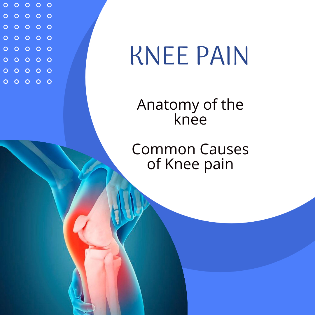
Knee Pain
The knee joint is one of the largest and most complex joints in the body. It is constructed by 4 bones and an extensive network of ligaments and muscles. The three main bones, the femur (thigh bone), tibia (shin bone) and the patella (knee cap) form the main articular surfaces, whereas the fourth bone on the outside of the lower leg known as the fibula mainly provides attachment for some of the muscles that move the knee and the lateral collateral ligament which helps stabilise the knee.
Between the femur and the tibia are 2 structures called ‘menisci’ (commonly referred to as cartilages). The menisci correct the lack of congruence between the articular surfaces of tibia and femur, increase the area of contact and improve weight distribution and shock absorption. They also help to guide and coordinate knee motion, making them very important stabilizers of the knee.
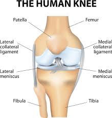
Within the knee joint are stabilising tissues known as ligaments. Each ligament has a particular function in helping to maintain optimal knee stability.
They are:
Anterior Cruciate
Posterior Cruciate
Medial Collateral
Lateral Collateral
There are also lots of muscles involved in both movement of the knee and stability. The main muscles are :
The Quadriceps
The Hamstrings
Gastrocnemius
Popliteus
Sartorius
Gracilis
Common Knee Injuries
There is an extensive list of cause of knee pain which can include
Ligament Injuries
Muscular Injuries
Tendinopathy
Meniscal Injury
Osgood Schlatters (Adolescence)
Bursitis
OA
Bakers Cyst
Patellofemoral Pain.
Assessment
A case history (questioning) can obtain an insight into possible causes of knee pain and you may be asked questions such as:
Any back or leg pain? (pain in the knee can be referred from the back)
Is there hip or ankle pain? (Knee pain can be referred from the hip or biomechanically affected by the ankle)
Does the knee give way? (instability/rupture of ligaments)
Does the knee lock? (meniscus, true locking associated with bucket handle tears)
Did the knee swell? How quickly? Where is the swelling? (Intra articular/ extra articular; immediate swelling usually indicates trauma within the knee such as ligament damage)
Was there bruising? (Immediate bruising indicates significant trauma)
Age – The following conditions are not exclusive to these age groups but a higher prevalence is noted in these populations (elderly – OA?, young – osgoods schlatters, middle aged- meniscal).
Type of shoes (wear patterns/age of shoes/proper design)
Examination
The osteopath will assess the movement of the knee actively and passively, weight bearing & non weight bearing as pain allows. Palpation for tender areas can give clues as to the site of pain as well as diagnostic/orthopaedic tests aimed at stressing different structures to elicit pain and aid the diagnosis. Signs of any muscle wasting or weakness can give valuable insight as well.
They will have a look at you walking to assess biomechanics or look for any altered gait due to pain.
Joints above and below the area will be looked at as well (ankle, hip, spine)
Treatment
In a majority of cases, knee pain can be managed conservatively which will include guidance on rest from sport, rehabilitation protocols and programme, strengthening advice.
For more information on recovery from ligament injury, follow the link below.
Occasionally further investigations are required and your osteopath should guide you accordingly.
